Valid Names Results
Paraputo martonoi Zarkani & Kaydan, 2025 (Pseudococcidae: Paraputo)Nomenclatural History
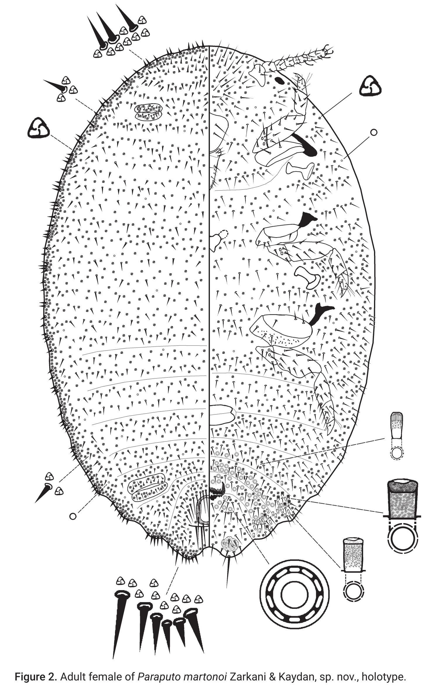
- Paraputo martonoi Zarkani & Kaydan 2025: 52. Type data: INDONESIA: East Kalimantan Province, Sepaku, on Rubiaceae, 11/15/2024, coll. A Zarkani. Holotype, female, by original designation Type depository: Bengkulu: Department of Plant Protection, Faculty of Agriculture, University of Bengkulu, Bengkulu, Indonesia; accepted valid name Notes: Paratypes: same data as holotypes, 3 ♀♀ on one slide, each slide with 2 specimens (MZB) Illustr.
Common Names
Ecological Associates
Hosts:
Families: 1 | Genera: 1
- Rubiaceae
- Rubiaceae | ZarkanReFa2025
Associates:
Families: 1 | Genera: 1
- Formicidae
- Dolichoderus | ZarkanReFa2025
Geographic Distribution
Countries: 1
- Indonesia
- Kalimantan (=Borneo) | ZarkanReFa2025
Keys
- ZarkanReFa2025: pp.63 ( Adult (F) ) [Paraputo found in Southeast and southern Asia (adapted from Williams (2004))]
Remarks
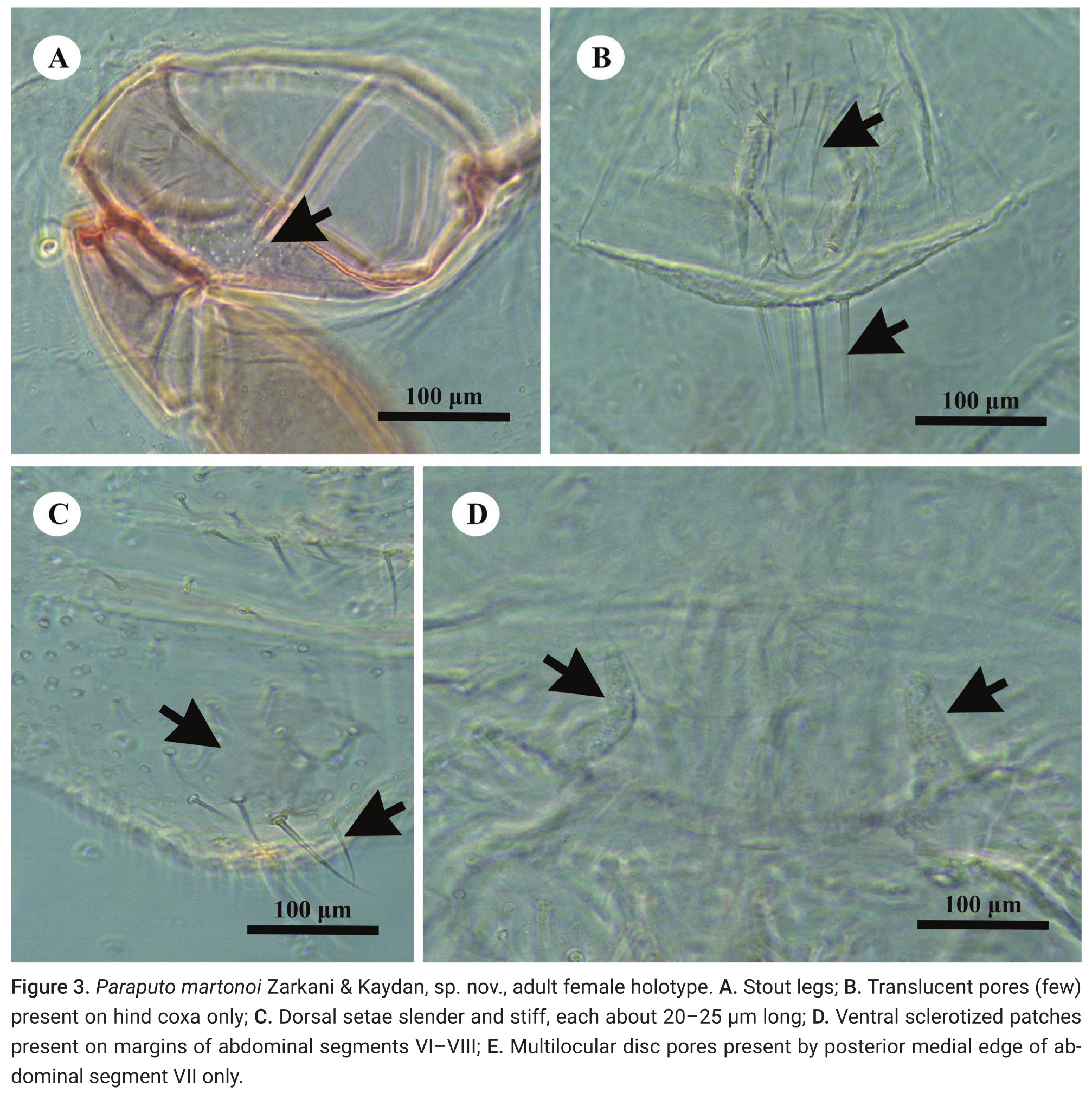
- Systematics: Paraputo martonoi is most similar to P. carnosae (Takahashi) in having: (i) large oral collar tubular ducts on abdomen only, each over 1.5 times as wide as a trilocular pore; (ii) a large circulus divided by an intersegmental line; (iii) setae on each side of anal ring, of similar length or a bit shorter than an anal ring seta, and ventral submarginal setae on abdominal segments V–VIII shorter than an anal ring seta; and (iv) ventral sclerotized patches present on anal lobes, also on ventral margins of abdominal segment VII, and sometimes VI. However, P. martonoi can be readily distinguished from P. carnosae by having: (i) dorsal surface with slender, stiff setae, each about 20–25 μm long; (ii) legs stout; (iii) translucent pores present on hind coxa only; (iv) oral collar tubular ducts present on abdominal segments VII and VIII; and (v) multilocular disc pores present by posterior medial edge of abdominal segment VII only. Paraputo martonoi also resembles P. latebrae Williams. (Zarkani et al. 2025)
- Structure: In life, adult females produce a powdery white wax covering the dorsal surface of the body. Slide-mounted adult female: Body broadly oval, sometimes almost rotund, membranous, largest specimens 2.13 mm long and 1.62 mm wide. Anal lobes weakly developed, each ventral surface bearing a stout apical seta 80 μm long arising from a large circular-to-oval sclerotized area, about 57–79 μm wide, on ventral margin of abdominal segment VIII; small sclerotized areas present also on ventral margins of segments VII and VI. Antennae each 340 μm in total length, with 7 antennomeres; antennal setae mostly short. Anterior spiracles each 110 μm long and 58 μm wide across the atrium, while the posterior spiracles are 110 μm long and 65 μm wide across the atrium. Legs well developed. (Zarkani et al. 2025)
- Biology: Living on woody parts of its host plant, commonly attended by ants of the genus Dolichoderus Lund (Zarkani et al. 2025).
- General Remarks: Description and illustration by Zarkani et al. (2025).
Illustrations
Citations
- ZarkanReFa2025: ant association, description, diagnosis, distribution, host, illustration, key, taxonomy, 52, 63