Valid Names Results
Aulacaspis buteae Takahashi, 1942 (Diaspididae: Aulacaspis)Nomenclatural History
- Aulacaspis buteae Takahashi 1942b: 37-38. Type data: THAILAND: Chiengmai, on Butea frondosa, 02/04/1940. Syntypes, female, Type depository: Taichung: Entomology Collection, Taiwan Agricultural Research Institute, Wu-feng, Taichung, Taiwan; accepted valid name Illustr.
Common Names
Ecological Associates
Hosts:
Families: 1 | Genera: 1
- Fabaceae
- Butea monosperma | Takaha1942b | (= Butea frondosa)
Geographic Distribution
Countries: 2
- Nepal | Takagi2018
- Thailand | Takaha1942b
Keys
Remarks
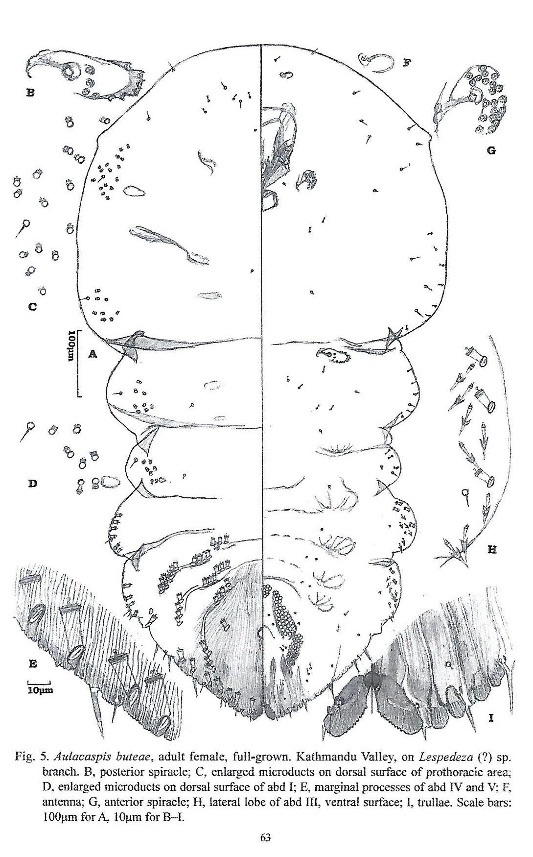
- Systematics: Aulacaspis buteae is related to A. rosae, but its median lobes are longer and more closely placed and the dorsal gland ducts are fewer on the pygidium. A. buteae can be told from A. tubercularis in the shape of the median lobes, the cephalothorax distinctly wider than long. It can be told from A. murrayae and A. phoebicola in lacking dorsal gland ducts on the basal abdominal segments (Takahashi, 1942b). A. buteae is known only from the ramicolous specimens. Takahashi makes no mention of the feeding site or sites of his specimens on Butea frondosa. It remains uncertain, therefore, whether the occurrence of enlarged microducts is a stable character in A. buteae. If the occurrence of enlarged microducts on the dorsal surface of the prosoma and the basal two segments of the postsoma is stable and constant in A. buteae, this character is helpful in recognizing the species. (Takagi, 2018)
- Structure: Adult female at full growth with prosoma well swollen, distinctly broader than postsoma, nearly quadrate or enlarged into an almost rounded mass; metathorax and abd II about the same in width, abd I narrower; pygidium nearly triangular. Prosomatic tubercles each represented by a small protuberance at most. Anterior spiracles each with a rather small loose cluster of pores; posterior spiracles each with some disc pores mostly arranged along a curved line, the lateralmost ones laid in a dermal fold. Enlarged dorsal microducts occurring submarginally on pro- and mesothoracic areas of prosoma, metathorax, and abd I, forming loose segmental clusters, the prothoracic cluster being distinctly separated from the mesothoracic one. (Takagi, 2018)
- General Remarks: Detailed description and illustration by Takahashi (1942b).
Illustrations
Citations
- Ali1969: distribution, host, taxonomy, 70
- Borchs1966: catalog, distribution, host, taxonomy, 136
- Scott1952: taxonomy, 35
- Takagi2018: description, diagnosis, distribution, illustration, 45-47, 63, 64
- Takaha1942b: description, distribution, host, illustration, taxonomy, 37-38


