Valid Names Results
Andaspis novaecaledoniae Hamilton & Williams, 2017 (Diaspididae: Andaspis)Nomenclatural History
- Andaspis novaecaledoniae Hamilton & Williams 2017: 25-27. Type data: NEW CALEDONIA: Rivière Bleue, Nothofagus codonandra, 10/10/1978. by J.S. Dugdale. Holotype, female, by original designation Type depository: London: The Natural History Museum, England, UK; accepted valid name Notes: Paratypes: 21 adult females. New Caledonia: Rivière Bleue and Mt. Mou. Collected on Nothofagus baumannae and N. codonandra, J.S. Dugdale and P.N. Johnson, 10./10/1978 and 11/2/1978. Deposited at BMNH and NMNH. Illustr.
Common Names
Ecological Associates
Hosts:
Families: 1 | Genera: 1
- Nothofagaceae
- Nothofagus baumanniae | HamiltWiHa2017
- Nothofagus codonandra | HamiltWiHa2017
Geographic Distribution
Countries: 1
- New Caledonia | HamiltWiHa2017
Keys
- HamiltWiHa2017: pp.30 ( Adult (F) ) [Andaspis from New Caledonia]
Remarks
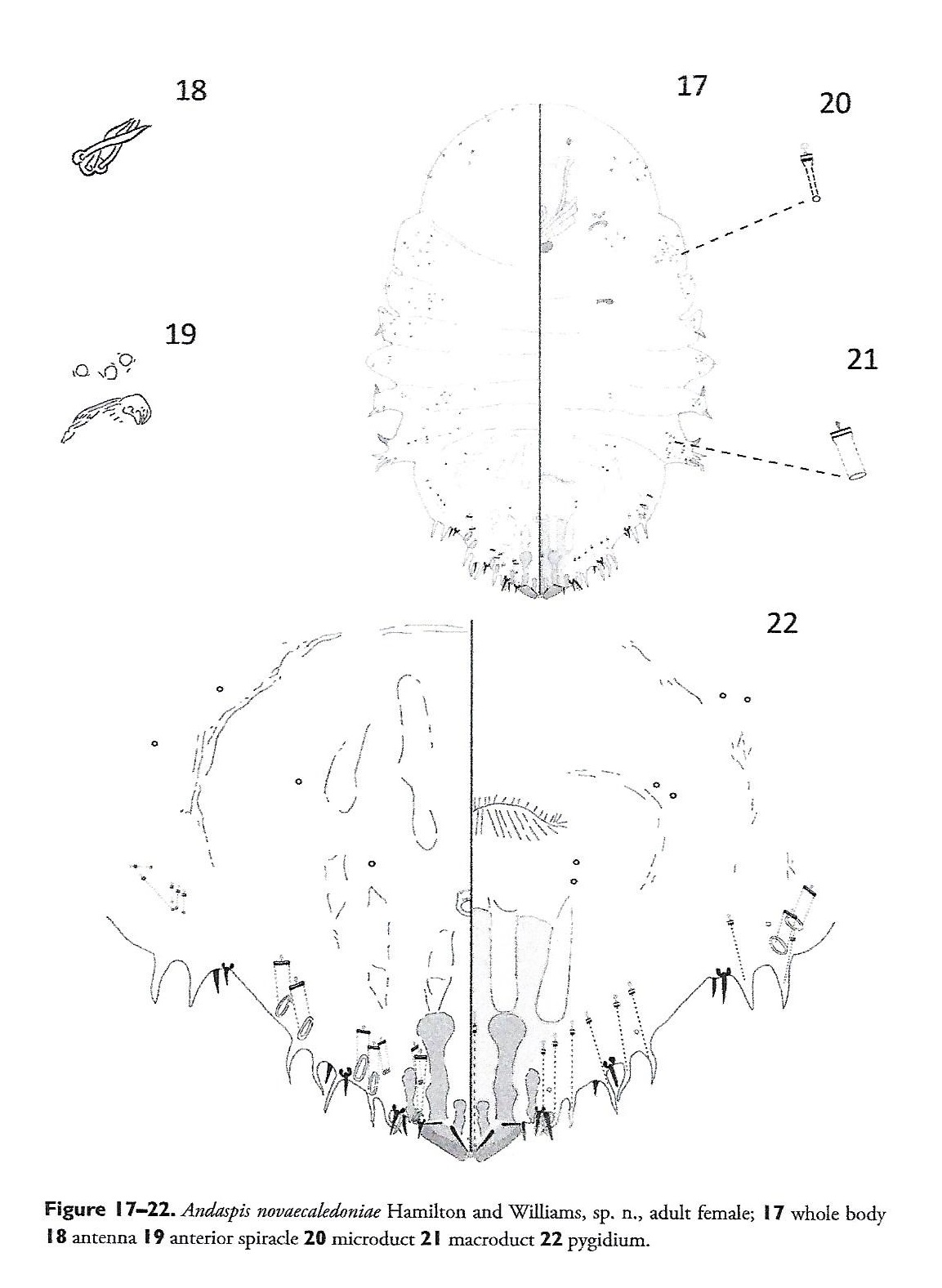
- Systematics: http://zoobank.org/5368CA1F-D60B-49FC-A36B-4BCB2BC8144C The adult female of this species is different from those of all other species in the genus described so far, in having two marginal macroducts located on the venter. Similarly, A. ornata sp. n. has nine marginal macroducts located on the venter. However, this species is smewhat similar to Andaspis tokyoensis Takagi and Kawai, 1966, a species known to occur in Japan. Adult females of A. novaecaledoniae and A. tokyoensis share well-developed lateral lobes on the abdomen, a club-shaped sclerosis arising from each median lobe, a narrow macroduct located on abdominal segment 7, and a sclerosis located anterolateral to each median lobe. This species differs from A. tokyoensis by the following characters (those for A. tokyoensis in parentheses): two scleroses located above each median lobe (one sclerosis located above each median lobe), five marginal macroducts and two submarginal macroducts located on the dorsum (six marginal macroducts and one submarginal macroduct located on the dorsum), lacking perivulvar pores (three groups of perivulvar pores), and antennae with three setae (antennae with two setae). (Hamilton, et al., 2017)
- Structure: Slide-mounted adult female 0.84–1.46 mm long; widest at first abdominal segment, 0.52–0.84 mm. Body outline oval or oblong, derm membranous except for pygidium. Each antenna with three setae. Anterior spiracles each with 1–4 disc pores, each about 5 μm in diameter, trilocular; posterior spiracles lacking pores. Anterior abdominal segments well-developed with convex margins; tooth-like tubercles present on segments 1, 3, and 4. In addition to those on pygidium, gland spines present along margins of abdominal segments 3 and 4. Many microducts distributed along margins and submargins of thorax and abdomen on both venter and dorsum, plus several on head. (Hamilton, et al., 2017)
- General Remarks: Detailed description and illustration in Hamilton, et al., 2017.
Illustrations
Citations
- HamiltWiHa2017: description, diagnosis, distribution, host, illustration, key, taxonomy, 25-27
- NormarOkMo2019: taxonomy, 58


